AP ELBOW
Anteroposterior projection protocol for elbow joint
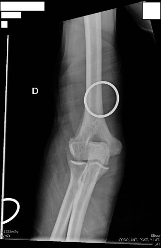
Important Note about Medical Devices
The rings visible in the radiograph correspond to a sling or arm sling
The immobilization device should not be removed until the full extent of the injury is observed on the radiographs.
This precaution is fundamental to avoid aggravating injuries during the radiological evaluation process.
Exposure Factors
Low exposure: Parameters optimized for elbow joint visualization
Anatomical Structures Visible
Should be clearly observed:
- Complete elbow joint
- Distal part of humerus (humeral condyles)
- Proximal part of ulna (olecranon and coronoid process)
- Proximal part of radius (radial head and neck)
- Joint spaces humeroulnar and humeroradial
- Medial and lateral epicondyles of humerus
Cassette Size and Division
Standard cassette for elbow
For multiple projections
Divided cassette: Allows performing AP and lateral projections on a single cassette for complete evaluation
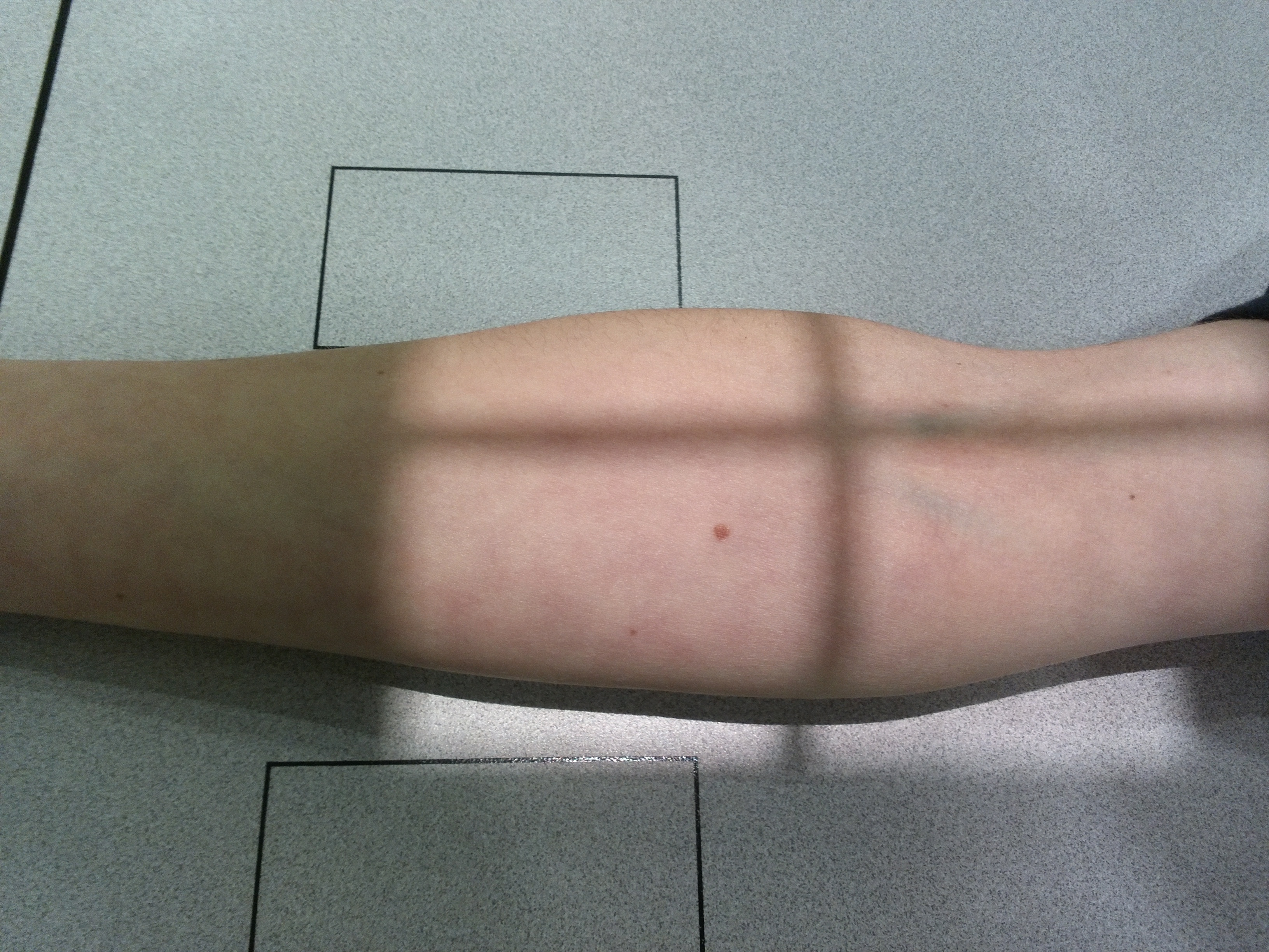
Patient Positioning
Central Ray Point
Direction: Vertical and perpendicular to center of joint
Location: Midpoint between humeral epicondyles
Goal: Humeroulnar joint space
Key Alignment Points
Extremity Alignment
• Arm and forearm in same plane
• Full elbow extension
• Hand supination
Epicondyles Parallel
• Medial and lateral epicondyles parallel to cassette
• Avoids humerus rotation
• Guarantees true AP view
Considerations for Injured Patients
Limited Extension
If full extension not possible:
• Extend to maximum tolerated
• Document degree of extension
• Consider alternative projection
Immobilization Devices
• Maintain sling during study
• Do not remove until complete evaluation
• Document presence in report
Patient Instructions
"Do not breathe during exposure"
Maintain position without movement during radiographic exposure
Special attention to keeping elbow extended and hand in supination
Optimal Image Characteristics
Complete joint
Distal humerus and proximal forearm
Without rotation
Epicondyles parallel to cassette
Joint spaces
Humeroulnar and humeroradial visible
Adequate field
Distal humerus to ulna/radius proximal
Common Technical Challenges
Frequent problems in AP elbow projection:
- Arm rotation causing oblique view
- Incomplete extension of elbow
- Insufficient supination of hand
- Poor alignment of epicondyles with cassette
- Structure superposition due to poor position
- Exclusion of joint parts due to poor centering
Solution: Verify that humeral epicondyles are parallel to cassette and maintain full extension with supination